Home

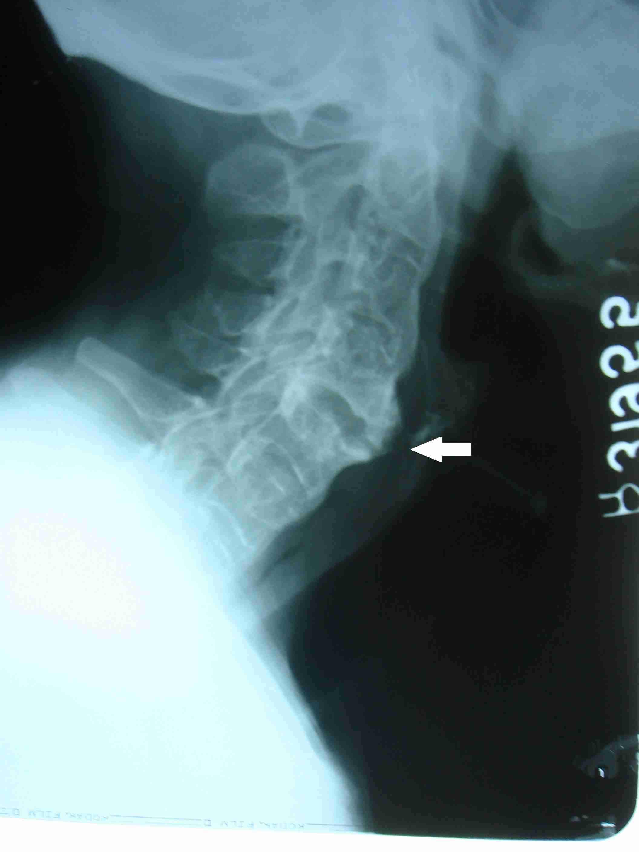
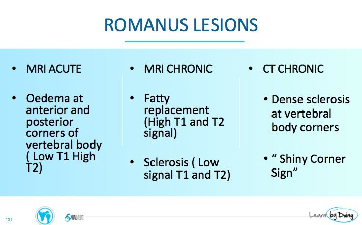
konversija vartojimas Grab romanus lesion prisukti pūtimo skylė vergas

Ankylosing spondylitis "Romanus lesions" | Radiology Case | Radiopaedia.org

The fatty Romanus lesion: a non-inflammatory spinal MRI lesion specific for  axial spondyloarthropathy | Annals of the Rheumatic Diseases
The fatty Romanus lesion: a non-inflammatory spinal MRI lesion specific for axial spondyloarthropathy | Annals of the Rheumatic Diseases
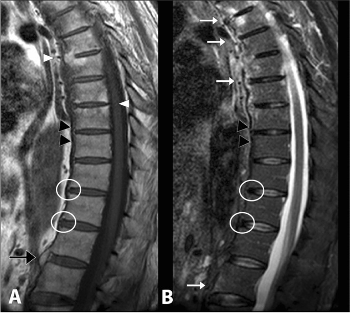
File:Romanus Lesion.webp - Physiopedia

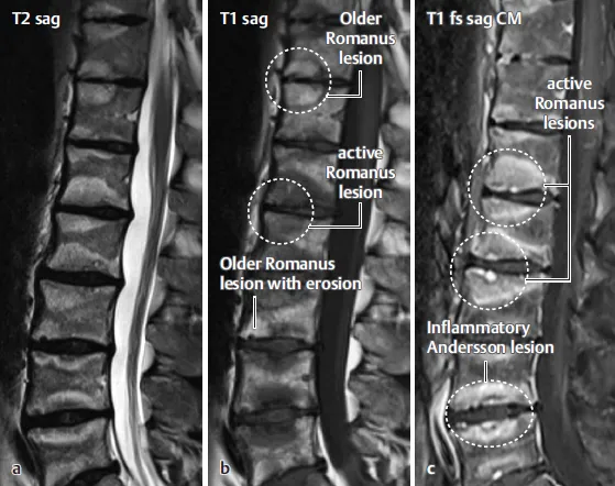
Differentiation between infectious spondylodiscitis versus inflammatory or  degenerative spinal changes: How can magnetic resonance imaging help the  clinician? | La radiologia medica
Differentiation between infectious spondylodiscitis versus inflammatory or degenerative spinal changes: How can magnetic resonance imaging help the clinician? | La radiologia medica

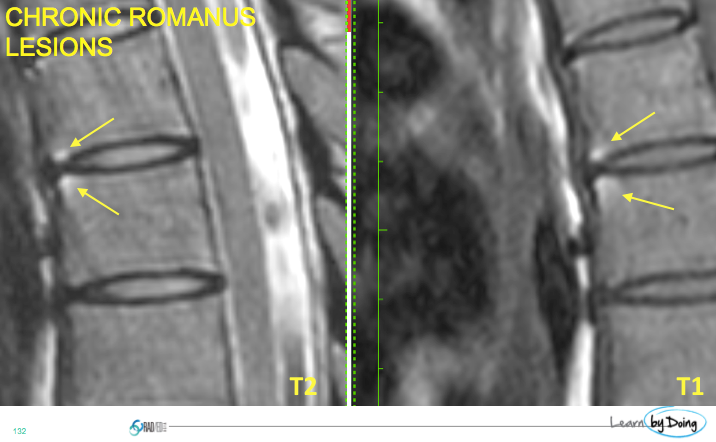
Old Romanus lesion. Sagittal T 1 weighted image | Download Scientific  Diagram
Old Romanus lesion. Sagittal T 1 weighted image | Download Scientific Diagram

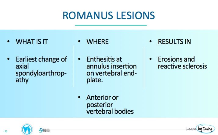
THE ROMANUS LESION: WHY, WHERE AND WHAT - Radedasia

EPOS™
EPOS™

Romanus lesions - ankylosing spondylitis | Radiology Case | Radiopaedia.org

Dr Tariq Tramboo on X: "When dealing with arthritis, do not forget the  spine. Spondyloarthritis; Structural lesions to look for include Romanus  lesion, Andersson lesion, syndesmophytes, hyperkyphosis and finally  ankylosis. https://t.co/4fekXIgyVw" /
Dr Tariq Tramboo on X: "When dealing with arthritis, do not forget the spine. Spondyloarthritis; Structural lesions to look for include Romanus lesion, Andersson lesion, syndesmophytes, hyperkyphosis and finally ankylosis. https://t.co/4fekXIgyVw" /

Beyond the sacro-Iliac joints: Vertebral involvement in axial  spondylarthritis - ScienceDirect
Beyond the sacro-Iliac joints: Vertebral involvement in axial spondylarthritis - ScienceDirect

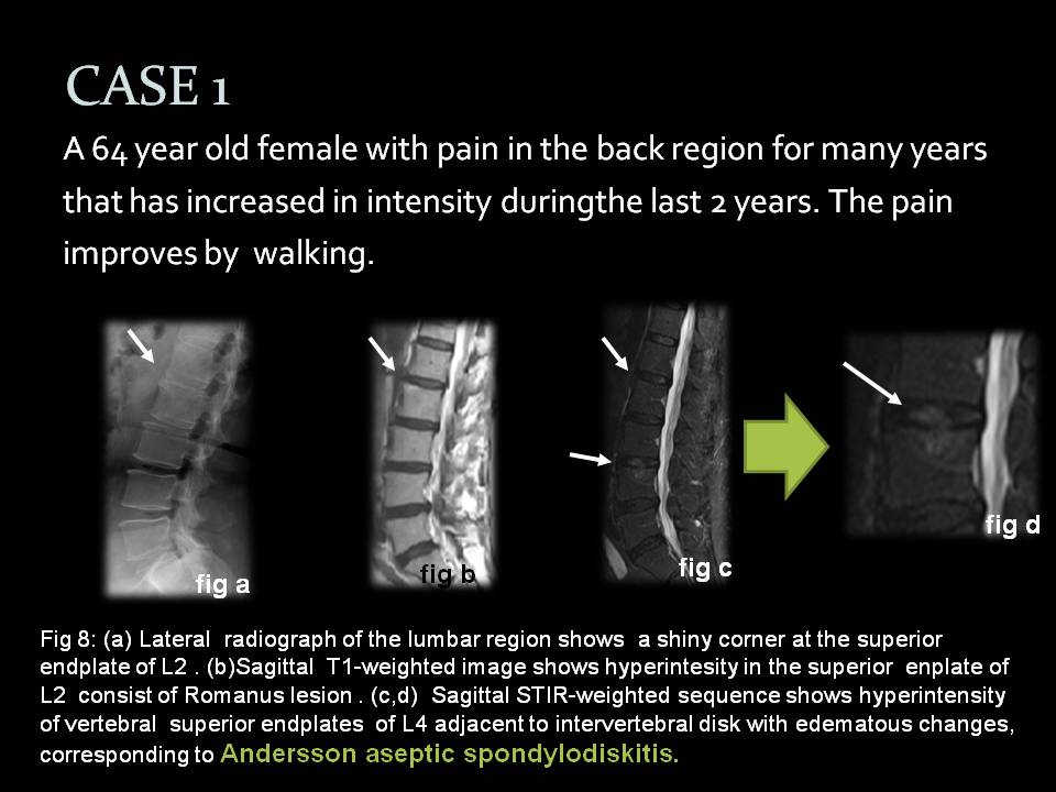
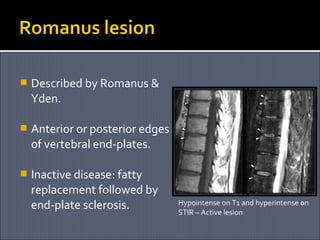
Ankylosing spondylitis | PPT

THE ROMANUS LESION: WHY, WHERE AND WHAT - Radedasia

THE ROMANUS LESION: WHY, WHERE AND WHAT - Radedasia

Ankylosing Spondylitis: MRI - Sumer's Radiology Blog

MR Corner Sign”: Value for Predicting Presence of Ankylosing Spondylitis

THE ROMANUS LESION: WHY, WHERE AND WHAT - Radedasia

Romanus lesions (MRI) | Radiology Case | Radiopaedia.org

The fatty Romanus lesion: a non-inflammatory spinal MRI lesion specific for  axial spondyloarthropathy | Annals of the Rheumatic Diseases
The fatty Romanus lesion: a non-inflammatory spinal MRI lesion specific for axial spondyloarthropathy | Annals of the Rheumatic Diseases

THE ROMANUS LESION: WHY, WHERE AND WHAT - Radedasia

Ankylosing spondylitis | PPT

JCDR - Ankylosing spondylitis, Sacroilitis, Bamboo spine, Romanus lesion,  Enthesitis
JCDR - Ankylosing spondylitis, Sacroilitis, Bamboo spine, Romanus lesion, Enthesitis

Figure 6 from Ankylosing spondylitis. | Semantic Scholar

Magnetic Resonance Imaging of Vertebral Erosion in Spondyloarthritis | The  Journal of Rheumatology
Magnetic Resonance Imaging of Vertebral Erosion in Spondyloarthritis | The Journal of Rheumatology

THE ROMANUS LESION: WHY, WHERE AND WHAT - Radedasia

Romanus lesion (vertebral bodies) | Radiology Reference Article |  Radiopaedia.org
Romanus lesion (vertebral bodies) | Radiology Reference Article | Radiopaedia.org

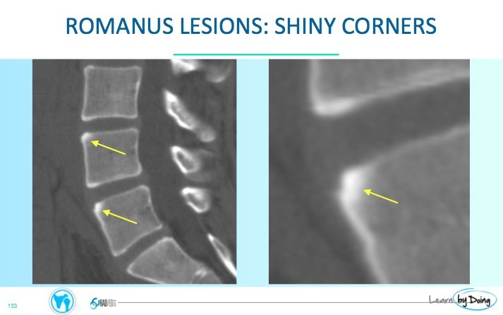
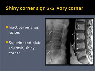
Romanus lesion shiny corner sign - NEET PG - www.MedicalTalk.Net the Best  Medical Forum for Medical Students and Doctors Worldwide
Romanus lesion shiny corner sign - NEET PG - www.MedicalTalk.Net the Best Medical Forum for Medical Students and Doctors Worldwide

Fatty corner lesions in T1-weighted magnetic resonance imaging as an  alternative to sacroiliitis for diagnosis of axial spondyloarthritis | BMC  Rheumatology | Full Text
Fatty corner lesions in T1-weighted magnetic resonance imaging as an alternative to sacroiliitis for diagnosis of axial spondyloarthritis | BMC Rheumatology | Full Text

EPOS™
EPOS™

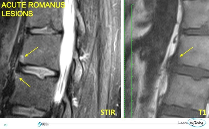
Spondylitis (active Romanus lesions) in a 34-year-old patient with... |  Download Scientific Diagram
Spondylitis (active Romanus lesions) in a 34-year-old patient with... | Download Scientific Diagram